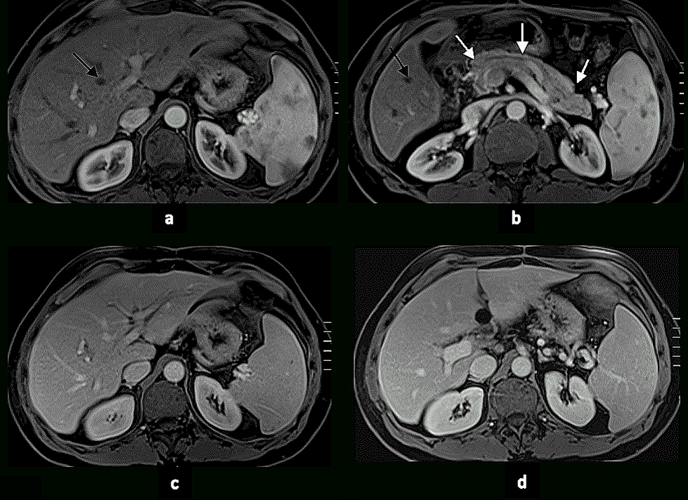
Figure 1:
Phases of Enhancement. T1-weighted post-gadolinium fat-suppressed hepatic arterial dominant phase (HADP)
(a, b), early hepatic venous phase (EHVP) (c) and interstitial phase (d) 3D-GE images at 1.5 T. Note that on the HADP (a, b) the contrast is present in the arteries (hepatic, renal, splenic and superior mesenteric); and renal, splenic, portal and superior mesenteric veins but not in the hepatic veins (black arrows a, b). The renal cortex demonstrates intense enhancement and the spleen, pancreas and liver demonstrate moderate enhancement. The moderate enhancement of the normal pancreas (white arrows b) reflects the adequate timing of enhancement of HADP. EHVP (c) shows enhancement of the entire vascular system of the liver and maximal enhancement of its parenchyma.
|